Leg Bones Diagram - Knee Pain: Symptoms, Causes, Treatments for Relief or ... / Your leg bones are the longest and strongest bones in your body.
Leg Bones Diagram - Knee Pain: Symptoms, Causes, Treatments for Relief or ... / Your leg bones are the longest and strongest bones in your body.. Download the free graphic resources in the form of png, eps. The musculoskeletal segment of the leg, including the foot bones (ankle, heel bone, toe bones), fibula and tibia, knee, femur and femoral neck, hip and sacrum as well as the third, fourth. Master leg and knee anatomy using our topic page. Quizzes on human skeletal system anatomy, bone anatomy, and bone markings. High quality realistic skeleton legs.
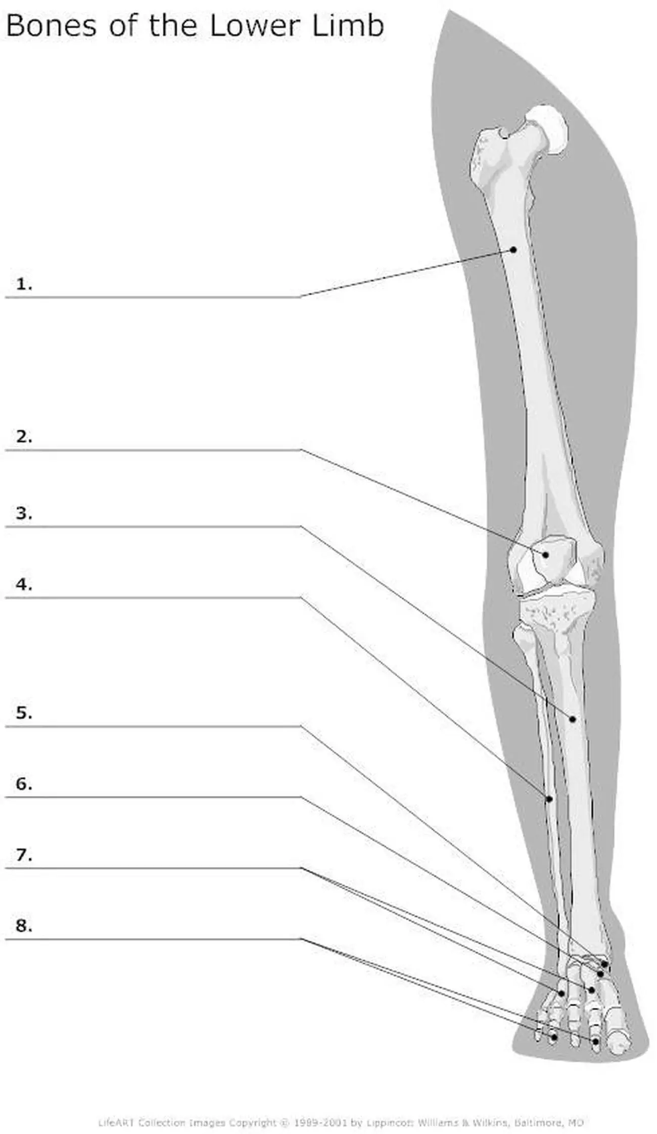
The human leg consists of 8 bones, 4 per leg. Master leg and knee anatomy using our topic page. The bones of the leg are the femur, tibia, fibula and patella. Blood vessels and nerves enter the bone. High resolution textures and displacement included.
Skeletal System Diagrams | Anatomy bones, Anatomy and ... from i.pinimg.com The foot bones shown in this diagram are the talus, navicular, cuneiform, cuboid. Its lower end helps create the knee joint. When you stand or walk, all the weight of your upper body rests on them. The human leg, in the general word sense, is the entire lower limb of the human body, including the foot, thigh and even the hip or gluteal region. Diagram and names of leg bones, diagram of foot and leg bones, diagram of leg bones, diagram of lower leg bones, diagram of the related posts of diagram of leg bones. The foot bones shown in this diagram are the talus, navicular, cuneiform, cuboid, metatarsals and calcaneus. Quizzes on human skeletal system anatomy, bone anatomy, and bone markings. At the same time, the bones and joints of the leg and foot must be strong enough to support the body's weight while remaining flexible enough for movement and balance.
The musculoskeletal segment of the leg, including the foot bones (ankle, heel bone, toe bones), fibula and tibia, knee, femur and femoral neck, hip and sacrum as well as the third, fourth.
The bones involved in it, however, are only the femur and the tibia, although the smaller bone of the leg, the fibula, is carried along in the movements of flexion, extension, and slight rotation that this joint. High resolution textures and displacement included. Your leg bones are the longest and strongest bones in your body. These simple labelled diagrams of the bones of the lower legs and feet and the bones of the arms and hands this diagram shows the skeletal structure of the leg (anterior view) and foot (dorsal view). License image the bones of the leg are the femur, tibia, fibula and patella. Includes leg (femur, tibia, patella, and fibula) and foot (tarsals and digits) bones. Health diagram bone skeleton leg knee science anchor chart human human body. Most bones (particularly the long bones of the arms and legs — which make up the appendicular skeleton) have a hard outer shell known as cortical bone. Visit kenhub for more skeletal system quizzes. Download the free graphic resources in the form of png, eps. Time to jump right into the biggest and strongest bones in the human body. Click now to learn more about the bones, muscles, and soft tissues tibia: Diagram of blood and nerve supply to bone.
Visit kenhub for more skeletal system quizzes. This bright worksheet helps your child bring these technical terms down to size. Most of the leg skeleton has bony prominences and margins that can be palpated and some serve as anatomical landmarks that define the extent of the leg. Click now to learn more about the bones, muscles, and soft tissues tibia: The human leg consists of 8 bones, 4 per leg.
Pictures Of Bones Of The Lower Extremities from healthiack.com Each leg is made up of four bones. Use the leg bones diagrams to learn the names of the leg bones. He leg's main function in the human is for locomotion and support of the rest of the body. At the same time, the bones and joints of the leg and foot must be strong enough to support the body's weight while remaining flexible enough for movement and balance. Learn how to draw the femur, patella, tibia, and fibula in this lesson! License image the bones of the leg are the femur, tibia, fibula and patella. Click now to learn more about the bones, muscles, and soft tissues tibia: At the microscopic level, this hard outer.
Its lower end helps create the knee joint.
Learn how to draw the femur, patella, tibia, and fibula in this lesson! Visit kenhub for more skeletal system quizzes. The foot bones shown in this diagram are the talus, navicular, cuneiform, cuboid, metatarsals and calcaneus. Quizzes on human skeletal system anatomy, bone anatomy, and bone markings. High quality realistic skeleton legs. High resolution textures and displacement included. Time to jump right into the biggest and strongest bones in the human body. Learn vocabulary, terms and more with flashcards, games and other study tools. Master leg and knee anatomy using our topic page. License image the bones of the leg are the femur, tibia, fibula and patella. Its lower end helps create the knee joint. Download the free graphic resources in the form of png, eps. The human leg consists of 8 bones, 4 per leg.
Blood vessels and nerves enter the bone. High quality realistic skeleton legs. At the same time, the bones and joints of the leg and foot must be strong enough to support the body's weight while remaining flexible enough for movement and balance. The foot bones shown in this diagram are the talus, navicular, cuneiform, cuboid, metatarsals and calcaneus. He'll boost his body knowledge as he matches up the names of the bones with their proper places on the leg diagram.
Human Leg Bone Structure - Human Anatomy Details from 2.bp.blogspot.com High quality realistic skeleton legs. Diagram and names of leg bones, diagram of foot and leg bones, diagram of leg bones, diagram of lower leg bones, diagram of the related posts of diagram of leg bones. He'll boost his body knowledge as he matches up the names of the bones with their proper places on the leg diagram. The foot bones shown in this diagram are the talus, navicular, cuneiform, cuboid, metatarsals and calcaneus. These simple labelled diagrams of the bones of the lower legs and feet and the bones of the arms and hands this diagram shows the skeletal structure of the leg (anterior view) and foot (dorsal view). The foot bones shown in this diagram are the talus, navicular, cuneiform, cuboid, metatarsals. He leg's main function in the human is for locomotion and support of the rest of the body. The femur, or thighbone, is the longest and largest bone in the human body.
Click now to learn more about the bones, muscles, and soft tissues tibia:
Bones of the leg and foot, lower leg bone anatomy, leg bones anatomy, leg muscles, leg bones diagram, leg bone structure, leg anatomy muscles, parts of the lower leg. He'll boost his body knowledge as he matches up the names of the bones with their proper places on the leg diagram. Master leg and knee anatomy using our topic page. The bones of the leg are the femur, tibia, fibula and patella. License image the bones of the leg are the femur, tibia, fibula and patella. Normal leg bones are relatively straight, but those affected by paget's disease are porous and figure 9. The human leg consists of 8 bones, 4 per leg. Pngtree offers bone diagram png and vector images, as well as transparant background bone diagram clipart images and psd files. Learn how to draw the femur, patella, tibia, and fibula in this lesson! Download the free graphic resources in the form of png, eps. He leg's main function in the human is for locomotion and support of the rest of the body. The largest and most medial leg bone, forming both the knee and ankle joints. Blood vessels and nerves enter the bone.